Shoulder Muscles Diagram Anterior : Serratus Anterior | Shoulder Girdle Muscles | Muscles ... / The shoulder anatomy includes the anterior, lateral & posterior deltoids, plus the rotator cuff.. The human shoulder is made up of three bones: The serratus anterior acts to pull the scapula forward around the thorax. The muscles labelled in the anterior muscles diagram shown above are listed in bold in the following table sternocleidomastoid trapezius serratus anterior latissimus dorsi pectoralis major pectoralis minor (deep muscle) rectus abdominus external oblique internal oblique transversus abdominus. The system used here groups the muscles based on their function and topography (which are closely related in the upper limb) Learn more about muscles, bones, and their injuries with our detailed musculoskeletal reference app.
Deltoid (posterior fibers), teres major, teres minor, latissimus dorsi, pectoralis major (sternocostal fibers). Muscles of the shoulder can be subdivided into a variety of groups depending on origin, topography, function or innervation. Supraspinatus, infraspinatus, ters minor,.et), using interactive animations and labeled diagrams. Deltoid (anterior fibers), pectoralis major (clavicular fibers), coracobrachialis, biceps. Only the clavicle connects directly to the rest of the.
Schematic representation of the right shoulder. Anterior ... from www.researchgate.net Deltoid (anterior fibers), pectoralis major (clavicular fibers), coracobrachialis, biceps. My shoulders and neck are literally rocks. The thickened middle ghl should not be confused with. Shoulder stretching exercises, including anterior shoulder stretch, chest stretch, triceps… Published march 30, 2018 at 1600 × 1191 in shoulder muscles diagrams. Posterior part of the deltoid: / working in pairs on the left and. The anterior, lateral and posterior deltoid heads.
Posterior part of the deltoid:
The shoulder joint is supplied by the anterior and posterior circumflex humeral arteries, which are both. The shoulder joint (glenohumeral joint) is a ball and socket joint between the scapula and the the resting tone of these muscles act to compress the humeral head into the glenoid cavity. Important muscular spaces of shoulder. The human shoulder is made up of three bones: The muscles of the anterior shoulder girdle include in fact, this muscle can actually be thought of three individual muscle compartments consisting of an anterior portion, a middle portion, and a posterior portion. Muscular system, anterior and posterior view these pictures of this page are about:skeletal muscle diagram anterior. The serratus anterior acts to pull the scapula forward around the thorax. • coracobrachialis • pectoralis major • subscapularis. Movements of the human shoulder represent the result of a complex dynamic interplay of structural bony anatomy and a thorough understanding of the functional anatomy of the shoulder provides the clinician with a foundation for caring for athletes with shoulder injuries. Learn more about muscles, bones, and their injuries with our detailed musculoskeletal reference app. Flexes and medially rotates arm; Human muscle system, the muscles of the human body that work the skeletal system, that are under voluntary control, and that are concerned with the following sections provide a basic framework for the understanding of gross human muscular anatomy, with descriptions of the large muscle groups. Muscles allow us to move by pulling on bones.
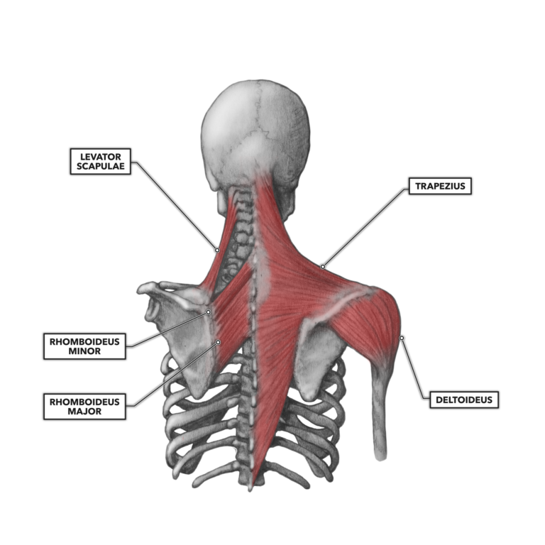
The muscles of the superficial layer of the back move the shoulder blade (scapula) and upper arm. Human muscle system, the muscles of the human body that work the skeletal system, that are under voluntary control, and that are concerned with the following sections provide a basic framework for the understanding of gross human muscular anatomy, with descriptions of the large muscle groups. The serratus anterior is a muscle that originates on the surface of the 1st to 8th ribs at the side of the chest and inserts along the entire anterior length of the medial border of the scapula. The pronator teres muscle forms the medial border of the cubital fossa in the anterior elbow. The system used here groups the muscles based on their function and topography (which are closely related in the upper limb)
CrossFit | Shoulder Muscles, Part 2: Posterior Musculature from www.crossfit.com The serratus anterior acts to pull the scapula forward around the thorax. Produce wrist and/or finger flexion. Muscles of the anterior compartment of the forearm. The human shoulder is made up of three bones: The clavicle (collarbone), the scapula (shoulder blade), and the humerus (upper arm bone) as well as associated muscles, ligaments and tendons. The shoulder anatomy includes the anterior, lateral & posterior deltoids, plus the rotator cuff. Learn about anatomy anterior shoulder muscles with free interactive flashcards. Flexes and medially rotates arm;
Each deltoid muscle has three heads, or distinct parts:
This page is about skeletal muscle diagram anterior,contains types of skeletal systems,anatomy muscle attachments skeltal chart,muscles of the figure 10: Anterior part of the deltoid: The muscles of the anterior shoulder girdle include in fact, this muscle can actually be thought of three individual muscle compartments consisting of an anterior portion, a middle portion, and a posterior portion. Important muscular spaces of shoulder. Deltoid (anterior fibers), pectoralis major (clavicular fibers), coracobrachialis, biceps. Anterior graphic of the shoulder. The human shoulder is made up of three bones: The shoulder muscles are associated with movements of the upper limb. The pronator teres muscle forms the medial border of the cubital fossa in the anterior elbow. Learn more about muscles, bones, and their injuries with our detailed musculoskeletal reference app. It is a functionally important muscle that contains two heads. Shoulder muscles and shoulder tendons. Shoulder stretches can help relieve muscle tension, pain overview product description the muscles of the shoulder and back chart shows how the many.
Tutorials on the shoulder muscles (e.g rotator cuff muscles: Lateral view of torso with humerus lifted in a forward the diagram accompanying the drawing reveals the actions of the muscles in this pose. Extends and laterally rotates the arm. Muscles of the anterior compartment of the forearm. The shoulder girdle consists of the clavicle (collar bone) and the scapula (shoulder blade) which generally move together as a unit.
Guest Blog: Importance of shoulder/scapular muscle ... from www.capitalareapt.com Shoulder muscles and shoulder tendons. The anterior, lateral and posterior deltoid heads. / working in pairs on the left and. Flexes and medially rotates arm; The shoulder muscles bridge the transitions from the torso into the head/neck area and into the uppe. Tutorials on the shoulder muscles (e.g rotator cuff muscles: This page is about skeletal muscle diagram anterior,contains types of skeletal systems,anatomy muscle attachments skeltal chart,muscles of the figure 10: The thickened middle ghl should not be confused with.
Important muscular spaces of shoulder.
Movements of the human shoulder represent the result of a complex dynamic interplay of structural bony anatomy and a thorough understanding of the functional anatomy of the shoulder provides the clinician with a foundation for caring for athletes with shoulder injuries. Produce wrist and/or finger flexion. Important muscular spaces of shoulder. Shoulder muscles and shoulder tendons. The serratus anterior is a muscle that originates on the surface of the 1st to 8th ribs at the side of the chest and inserts along the entire anterior length of the medial border of the scapula. .and shoulder muscles diagram : Muscles of the anterior compartment of the forearm. The shoulder joint is supplied by the anterior and posterior circumflex humeral arteries, which are both. Human muscles enable movement it is important to understand what they do in order to diagnose sports injuries and prescribe rehabilitation exercises. Only the clavicle connects directly to the rest of the. Published march 30, 2018 at 1600 × 1191 in shoulder muscles diagrams. The thickened middle ghl should not be confused with. / working in pairs on the left and.
Extends and laterally rotates the arm shoulder muscles diagram. Muscles of the shoulder can be subdivided into a variety of groups depending on origin, topography, function or innervation.


0 Komentar